
You might have noticed tiny openings or pits along the edge of your eyelids—especially near the lashes—and wondered if they’re normal. These small indentations, often mistaken for abnormalities, are actually part of your eye’s natural anatomy. While most are harmless, some changes can signal underlying conditions. Understanding what these “holes” are, why they exist, and when to seek medical attention is essential for maintaining ocular health.
What Are the Holes in Your Eyelids?
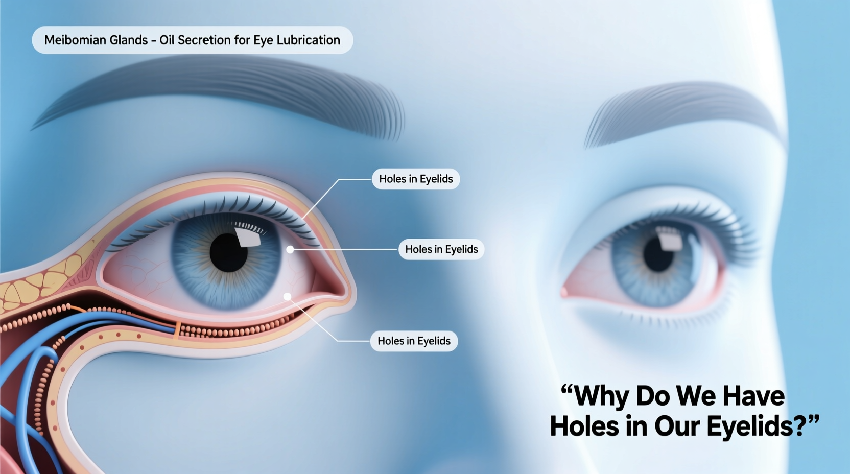
The so-called \"holes\" in your eyelids are typically one of two anatomical features: puncta or sebaceous gland openings. Neither are defects—they are functional parts of your eye's drainage and lubrication system.
The most noticeable are the puncta, which are tiny openings located on the inner corners of both the upper and lower eyelids, right next to the tear ducts. Each eyelid has one punctum, making four total per person. These act as entry points into the tear drainage system. When you blink, tears move across the eye surface and collect near the nose, where they enter through the puncta and travel down the nasolacrimal duct into the nasal cavity.
Another type of small opening includes the orifices of Meibomian glands, which line the inside edge of each eyelid. These glands secrete an oily substance that prevents tears from evaporating too quickly. Their openings can sometimes appear as minute dots or pits, especially under close inspection.
Why Do Puncta Exist? The Role of Tear Drainage
Tears aren’t just emotional responses; they play a crucial role in eye health by lubricating the surface, washing away debris, and protecting against infection. But constant tearing would flood the eyes without an efficient drainage system.
The puncta serve as the starting point of this system. Here’s how it works:
- Tears spread across the eye with each blink.
- They collect in the medial corner (near the nose) of the eye. <3>Fluid enters the puncta and moves into the canaliculi—small channels leading to the lacrimal sac.
- From there, tears flow down the nasolacrimal duct into the back of the nose and throat, where they’re swallowed unnoticed.
This continuous cycle keeps the eyes moist while preventing overflow. If the puncta become blocked, however, excessive tearing (epiphora) may occur—even in dry environments.
When Should You Be Concerned?
In most cases, visible puncta or gland openings are completely normal. However, certain symptoms accompanying these structures warrant medical evaluation:
- Persistent watery eyes – Could indicate a blocked tear duct.
- Swelling or redness near the inner eye – May suggest infection like dacryocystitis.
- Yellow or green discharge – A sign of bacterial infection.
- Pain or tenderness below the inner corner of the eye – Often linked to duct obstruction.
- Crusting along eyelid margins – Common in Meibomian gland dysfunction.
“While puncta are a normal feature, persistent tearing or recurrent infections should never be ignored. Early diagnosis can prevent complications like chronic sinus issues or corneal irritation.” — Dr. Lena Torres, Oculoplastic Surgeon
Common Conditions Related to Eyelid Openings
Several disorders affect the function or appearance of these tiny but vital structures. Below is a summary of key conditions:
| Condition | Description | Symptoms | Treatment |
|---|---|---|---|
| Nasolacrimal Duct Obstruction | Blockage in the tear drainage pathway beyond the puncta | Watery eyes, mucus discharge, frequent eye infections | Massage, probing, stenting, or surgery (dacryocystorhinostomy) |
| Meibomian Gland Dysfunction (MGD) | Chronic blockage of oil glands in eyelids | Dry eyes, gritty sensation, crusting upon waking | Warm compresses, lid hygiene, oral antibiotics, LipiFlow therapy |
| Punctal Stenosis | Narrowing or closure of the punctal opening | Excessive tearing, discomfort in wind or cold | Dilation, intubation, or surgical correction |
| Blepharitis | Inflammation of eyelid margins affecting gland openings | Redness, flaking, burning, blurred vision | Cleansing routines, anti-inflammatory drops, antibiotic ointments |
Mini Case Study: Recurrent Eye Watering in a 45-Year-Old Office Worker
Sarah, a 45-year-old administrative assistant, began experiencing constant tearing in her left eye over several months. She initially assumed allergies were to blame and used antihistamine eye drops without improvement. Upon visiting an ophthalmologist, she was diagnosed with partial nasolacrimal duct obstruction. Her puncta were visible and intact, but a dye disappearance test showed poor drainage. After undergoing a minimally invasive probing procedure, her symptoms resolved within a week. This case highlights how structural issues behind normal-looking “holes” can still impair function.
How to Maintain Healthy Tear Drainage and Gland Function
Maintaining the health of your eyelid openings doesn't require complex routines, but consistency matters. Follow this checklist to support optimal eye surface balance:
- Apply warm compresses for 5–10 minutes daily to keep Meibomian glands clear.
- Gently massage eyelids in a circular motion after warming to express oils.
- Use a mild cleanser (like diluted baby shampoo) on a clean cloth to wipe eyelid margins.
- Avoid rubbing eyes, especially with unwashed hands.
- Stay hydrated and consider omega-3 supplements to improve tear quality.
- Protect eyes from wind and dry air with wraparound sunglasses.
Step-by-Step Guide to Performing a Warm Compress for Eyelid Health
Blocked glands contribute to dryness and inflammation. A simple warm compress helps melt thickened oils and restore flow.
- Wash your hands thoroughly with soap and water.
- Soak a clean washcloth in warm (not hot) water. Wring out excess moisture.
- Close your eyes and place the cloth over both eyelids for 5–10 minutes.
- Re-wet the cloth as needed to maintain warmth.
- After removal, gently massage the outer and inner portions of the eyelids using your fingertip or a cotton swab.
- Follow with lid cleansing if recommended by your doctor.
Perform this routine once or twice daily, particularly if you suffer from dry eye symptoms or blepharitis.
Frequently Asked Questions
Are holes in the eyelids a birth defect?
No. The puncta are normal anatomical structures present in everyone. They form during fetal development as part of the tear drainage system. True congenital abnormalities—such as absent puncta or complete duct atresia—are rare and usually accompanied by severe tearing from infancy.
Can you see the puncta without a mirror?
Most people can see their puncta using a magnifying mirror. They appear as small black or dark gray dots on the inner rim of the eyelid, closest to the nose. If you wear glasses or contacts, your eye care provider may point them out during an exam.
Is it safe to touch or clean the puncta?
No. You should never insert anything into the puncta. Cleaning around the eyelid margin is acceptable, but direct manipulation can cause injury, scarring, or infection. Leave clinical procedures to trained professionals.
Conclusion: Know Your Anatomy, Protect Your Vision
The tiny holes in your eyelids are not flaws—they’re critical components of a finely tuned system that keeps your eyes comfortable and healthy. Recognizing what’s normal versus what requires attention empowers you to take proactive steps in eye care. Whether it’s managing dryness, addressing chronic tearing, or simply understanding your body better, knowledge is the first line of defense.


 浙公网安备
33010002000092号
浙公网安备
33010002000092号 浙B2-20120091-4
Comments
No comments yet. Why don't you start the discussion?