
A positron emission tomography (PET) scan is a powerful diagnostic imaging technique that provides detailed insights into how your body’s tissues and organs are functioning at a cellular level. Unlike standard imaging tests such as X-rays, CT scans, or MRIs—which primarily show structure—a PET scan reveals metabolic activity. This makes it especially valuable in detecting early-stage diseases, monitoring treatment effectiveness, and guiding medical decisions.
Used frequently in oncology, cardiology, and neurology, PET scans help physicians identify abnormalities before structural changes become visible. If you or a loved one has been recommended for a PET scan, understanding why it's done, what happens during the procedure, and how it works can ease anxiety and improve preparedness.
Why a PET Scan Is Done
PET scans are performed to evaluate organ and tissue function. Because they measure biological processes such as glucose metabolism, blood flow, and oxygen use, they offer a dynamic view of how well different parts of the body are working.
The most common reasons for undergoing a PET scan include:
- Cancer detection and staging: PET scans can locate tumors, determine whether cancer has spread, assess response to therapy, and detect recurrence.
- Heart disease evaluation: They help identify areas of the heart with reduced blood flow or damaged tissue, particularly in patients being considered for revascularization procedures.
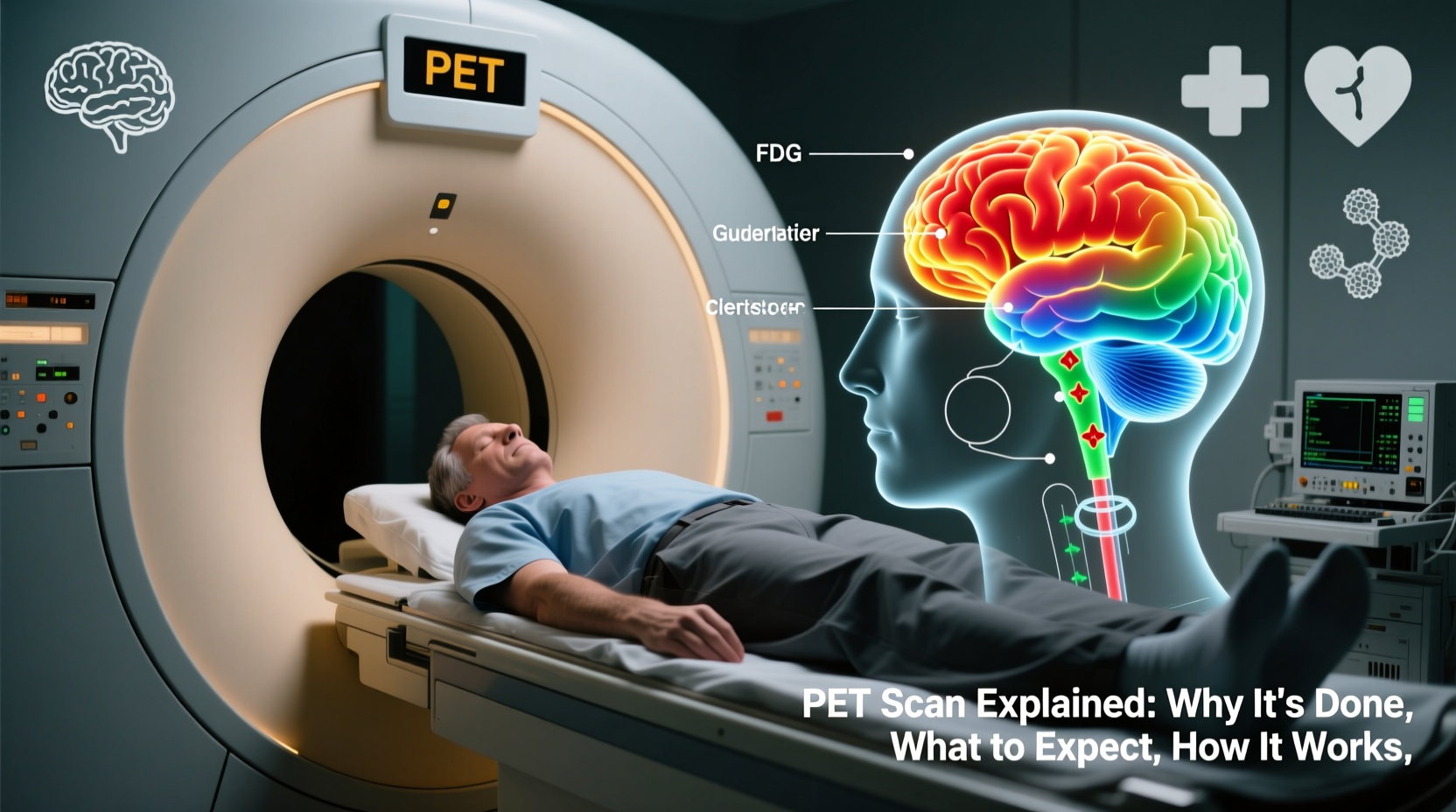
- Brain disorders: Neurological conditions like Alzheimer’s disease, epilepsy, and Parkinson’s can be assessed by observing patterns of brain metabolism.
- Infection or inflammation detection: In complex cases, PET scans can pinpoint hidden infections, such as those involving prosthetic joints or endocarditis.
“PET imaging allows us to see disease in action. It’s not just about where a tumor is—it’s about how active it is.” — Dr. Lena Patel, Nuclear Medicine Specialist
How a PET Scan Works
The science behind a PET scan lies in nuclear medicine. The procedure uses a radioactive substance called a radiotracer—most commonly fluorodeoxyglucose (FDG), a modified form of glucose. Since rapidly dividing cells (like cancer cells) consume more glucose than normal cells, they absorb higher amounts of the tracer.
Here’s how the process unfolds:
- The radiotracer is injected into a vein, usually in the arm.
- Over the next 30 to 60 minutes, the patient rests quietly while the tracer distributes throughout the body and accumulates in metabolically active tissues.
- The patient lies on a table that slides into the PET scanner, which detects gamma rays emitted when positrons from the tracer collide with electrons in the body.
- A computer converts this data into 3D images that highlight areas of high metabolic activity—often referred to as “hot spots.”
These images allow doctors to distinguish between benign and malignant growths, assess organ viability, and monitor biochemical changes over time. When combined with a CT scan (in a PET/CT), the results provide both functional and anatomical detail, increasing diagnostic accuracy.
What to Expect During the Procedure
Knowing what happens step by step can reduce anxiety and help you prepare mentally and physically.
Preparation
You’ll typically be asked to fast for 4–6 hours before the scan to stabilize blood sugar levels, which influence tracer uptake. Diabetic patients receive special instructions to manage their glucose. You should also avoid caffeine and intense physical activity the day before.
Upon arrival, you may be asked to change into a hospital gown and remove metal objects. A healthcare provider will inject the radiotracer intravenously. Then, you’ll rest in a quiet room for about an hour to allow the tracer to circulate.
During the Scan
When it’s time for imaging, you’ll lie on a motorized table that moves slowly through the donut-shaped scanner. It’s important to remain still during the 30–60 minute scan to ensure clear images. The machine doesn’t touch you, and the process is painless. Some people feel slightly claustrophobic, but the open design of most scanners helps minimize this.
After the Scan
Once complete, you can usually resume normal activities. The radioactive tracer decays quickly and is mostly eliminated through urine within a few hours. Drinking extra water helps flush it from your system faster. Results are interpreted by a nuclear medicine physician and sent to your doctor within a few days.
Do’s and Don’ts Before a PET Scan
| Do’s | Don’ts |
|---|---|
| Fast as instructed (usually 4–6 hours) | Eat or drink anything except water during fasting period |
| Stay hydrated after the scan | Engage in vigorous exercise 24 hours prior |
| Inform staff if you’re pregnant or breastfeeding | Wear clothing with metal zippers or jewelry |
| Bring a list of current medications | Consume sugary foods or drinks before the scan |
| Arrive early to complete paperwork | Drive immediately after if sedated (rarely needed) |
Real-World Example: Detecting Lung Cancer Early
Consider the case of Mark T., a 58-year-old former smoker. A routine chest X-ray revealed a small nodule in his lung. While CT imaging showed its size and location, it couldn’t confirm whether it was cancerous. His doctor ordered a PET scan. The results showed high metabolic activity in the nodule—indicating malignancy—while surrounding lymph nodes remained inactive. This allowed surgeons to proceed confidently with a targeted resection. Post-surgery pathology confirmed non-small cell lung cancer, caught early thanks to the PET scan. Mark began treatment promptly and remains in remission two years later.
This scenario illustrates how PET scans add critical value beyond structural imaging—offering functional insight that directly impacts diagnosis and treatment planning.
Frequently Asked Questions
Is a PET scan safe?
Yes, PET scans are generally safe. The amount of radiation exposure is low and comparable to other imaging tests like CT scans. The radiotracer has a short half-life and leaves the body quickly. Allergic reactions are extremely rare.
Will I feel anything during the scan?
No. The injection may cause a brief pinch, but the scanning process itself is painless. You won’t feel the radiation. Some find lying still for up to an hour uncomfortable, but most tolerate it well.
Can a PET scan diagnose Alzheimer’s disease?
It can support the diagnosis. Special tracers bind to amyloid plaques in the brain, which are associated with Alzheimer’s. When combined with clinical evaluation, PET imaging improves diagnostic confidence, especially in early or unclear cases.
Final Thoughts and Next Steps
A PET scan is more than just another test—it’s a window into the inner workings of your body. Whether you're being evaluated for cancer, heart disease, or a neurological condition, the information it provides can be pivotal in shaping your care plan. Understanding the purpose, process, and practical details helps you approach the scan with clarity and confidence.


 浙公网安备
33010002000092号
浙公网安备
33010002000092号 浙B2-20120091-4
Comments
No comments yet. Why don't you start the discussion?