
X-rays are one of the most common and valuable tools in modern medicine, used billions of times each year to diagnose fractures, infections, tumors, and other internal conditions. If you've ever had an X-ray, you’ve likely seen the striking image of your skeleton—white bones sharply defined against a gray or black background. But why do X-rays show bones so clearly while soft tissues appear faint or nearly invisible? The answer lies in physics, biology, and the way different materials interact with high-energy radiation.
How X-Rays Work: A Brief Overview
X-rays are a form of electromagnetic radiation, similar to visible light but with much higher energy and shorter wavelengths. This allows them to penetrate through many materials, including human tissue. When an X-ray machine is activated, it emits a controlled beam of X-ray photons that pass through the body and strike a detector on the other side.
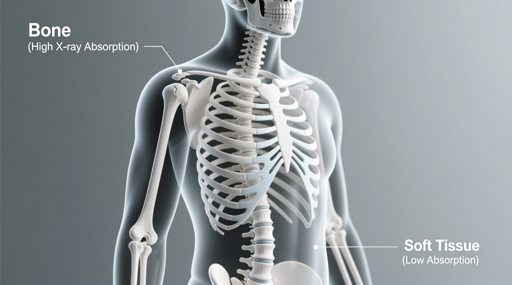
The resulting image is created based on how much radiation reaches the detector. Tissues that absorb more X-rays—like bone—block the photons from reaching the detector, appearing white or light gray. Softer tissues, such as muscle or fat, allow more rays to pass through, creating darker areas on the image.
Why Bones Appear White: The Role of Density and Calcium
Bones stand out so vividly in X-ray images because they are dense and rich in calcium, an element with a relatively high atomic number (20). Elements with higher atomic numbers have more electrons, which increases their ability to absorb X-ray photons through a process called photoelectric absorption.
When X-rays encounter bone, a significant portion of the beam is absorbed or scattered. As a result, fewer photons reach the detector behind the bone, leading to a lighter area on the image. In contrast, soft tissues like lungs, skin, or intestines are made mostly of lighter elements—carbon, hydrogen, oxygen, and nitrogen—which have low atomic numbers and weak interactions with X-rays. These tissues absorb far less radiation, allowing most photons to pass through and create a darker appearance on the film.
“Bone’s mineral matrix, especially hydroxyapatite containing calcium and phosphate, makes it exceptionally effective at attenuating X-rays compared to surrounding soft tissues.” — Dr. Alan Reeves, Radiological Physicist, Massachusetts General Hospital
Comparing Tissue Types: What Shows Up and Why
Not all body tissues interact with X-rays in the same way. Their visibility depends on three main factors: density, thickness, and atomic composition. Below is a comparison of common tissue types and their X-ray visibility.
| Tissue Type | Density Level | Atomic Composition | Appearance on X-Ray |
|---|---|---|---|
| Bone | Very High | Calcium, Phosphorus | White/light gray |
| Muscle | Moderate | Carbon, Oxygen, Hydrogen | Medium gray |
| Fat | Low | Hydrocarbons | Dark gray |
| Lung Tissue | Very Low (air-filled) | Gas + soft tissue | Black/dark |
| Cartilage | Low-Moderate | Collagen, water | Faint, often not visible |
This differential absorption is what creates contrast in X-ray imaging. Without variation in tissue density, the image would be uniformly gray, making diagnosis impossible.
Limitations of Standard X-Rays and Enhancing Visibility
While X-rays excel at visualizing bones, they have limitations when it comes to soft tissues. For example, ligaments, tendons, spinal discs, and brain tissue are rarely visible on conventional X-rays. To overcome this, radiologists sometimes use contrast agents—substances introduced into the body to increase the visibility of specific structures.
For instance, barium sulfate is swallowed or administered rectally to highlight the digestive tract, while iodine-based dyes can illuminate blood vessels or the urinary system. These agents contain high-atomic-number elements that strongly absorb X-rays, making hollow or fluid-filled organs suddenly visible.
Step-by-Step: How an X-Ray Image Is Formed
- X-ray generation: The machine produces a focused beam of X-ray photons.
- Beam penetration: Photons pass through the patient’s body.
- Differential absorption: Dense tissues like bone absorb more photons; soft tissues allow more to pass.
- Detection: Remaining photons hit a digital sensor or photographic film.
- Image formation: Areas with fewer photons (blocked by bone) appear lighter; areas with more photons appear darker.
- Interpretation: A radiologist analyzes the contrast patterns to identify abnormalities.
Real-World Example: Diagnosing a Wrist Fracture
Sarah, a 28-year-old cyclist, fell during a trail ride and landed on her outstretched hand. At the emergency clinic, she complained of pain and swelling in her wrist. A standard X-ray was ordered.
The resulting image showed a clean, white outline of the forearm bones (radius and ulna), wrist bones (carpals), and hand bones (metacarpals and phalanges). However, one of the small carpal bones—the scaphoid—showed a thin, dark line running through its normally solid white structure. This indicated a fracture, as the break allowed more X-rays to pass through at that point.
Because the scaphoid has a poor blood supply and is prone to non-healing, early detection via X-ray was crucial. Sarah was placed in a cast and referred for follow-up imaging. This case illustrates how even subtle changes in bone density captured by X-rays can guide life-impacting treatment decisions.
Common Misconceptions About X-Ray Imaging
- Misconception: X-rays \"light up\" bones like a flashlight. Reality: Bones appear bright because they block the light (X-rays), not because they emit it.
- Misconception: All body parts show up equally well on X-rays. Reality: Only structures with sufficient density or contrast are visible without enhancement.
- Misconception: X-rays use reflection, like mirrors. Reality: X-ray imaging relies on transmission—measuring what passes through, not what bounces back.
Frequently Asked Questions
Can X-rays show muscles or ligaments?
No, standard X-rays cannot clearly show muscles, ligaments, or tendons because these soft tissues lack the density and atomic composition needed to absorb enough X-rays for contrast. MRI or ultrasound is better suited for evaluating soft tissue injuries.
Why do teeth also appear white on X-rays?
Teeth, like bones, are rich in calcium and minerals such as enamel and dentin, which are highly radio-opaque. This makes them appear bright white on dental X-rays, allowing dentists to detect cavities, abscesses, and root issues.
Are X-rays harmful because they pass through the body?
While X-rays are ionizing radiation and carry some risk, diagnostic doses are very low and tightly regulated. The benefits of accurate diagnosis typically outweigh the minimal risk. Pregnant women and children receive special consideration to minimize exposure.
Practical Tips for Patients Undergoing X-Rays
- Wear loose, comfortable clothing
- Remove metal accessories (watches, necklaces, zippers)
- Inform staff about pregnancy or implanted devices
- Follow instructions to stay still during exposure
- Ask for a copy of your images for personal records
Conclusion: Seeing the Invisible Through Science
The reason X-rays show bones so clearly is a perfect intersection of physics and biology. Bone’s high density and calcium content make it a natural shield against X-ray photons, creating the contrast necessary for clear imaging. While soft tissues fade into the background, bones emerge in sharp relief—providing clinicians with vital information for diagnosis and treatment.
Understanding this principle not only demystifies a common medical procedure but also highlights the elegance of using physical laws to peer inside the human body non-invasively. Whether confirming a broken arm or screening for disease, X-ray technology remains a cornerstone of healthcare because it turns the invisible into the visible—one photon at a time.


 浙公网安备
33010002000092号
浙公网安备
33010002000092号 浙B2-20120091-4
Comments
No comments yet. Why don't you start the discussion?