
All categories
Featured selections
Trade Assurance
Buyer Central
Help Center
Get the app
Become a supplier
Customization:
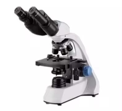
The Competitive Price Binocular Biological Microscope for Training is a robust, versatile optical instrument designed for educational and research environments. Featuring magnifications up to 2500x, it combines durability (metal and durable plastic build) with ergonomic design, enabling precise observation across diverse applications like biology, chemistry, and materials science.
| Feature | Specification | Application Scenario |
|---|---|---|
| Material | High-quality metal and durable plastics | Withstands frequent use in labs and classrooms |
| Magnification | 100x, 1000x, 2500x (via objectives) | Detailed analysis of cells, tissues, and microstructures |
| Eyepiece | Adjustable 10x binocular | Comfortable viewing during prolonged use |
| Stage | Mechanical, adjustable positioning | Precise sample alignment for advanced techniques (e.g., phase contrast) |
| Light Source | LED illumination (bright/darkfield) | Versatile lighting for diverse sample types |
Adjustable magnification levels (100x–2500x) and mechanical stage positioning allow customization to meet specific training or research needs, such as optimizing focus for delicate samples or enhancing visibility in low-light conditions.
Ideal for students and researchers, this microscope bridges affordability and performance. Its high magnification and ergonomic design make it perfect for labs, classrooms, or field studies, enabling clear observation of microscopic details without compromising durability.
| Parameter | Base Model | Advanced Model | Pro Model |
|---|---|---|---|
| Max Magnification | 1000x | 2500x | 2500x (+15% resolution) |
| Resolution | 0.8 NA | 1.0 NA | 1.2 NA |
| Light Intensity | 800 lux | 1000 lux | 1200 lux |
Technical Breakthroughs:
Version Selection Guidance:
With the Pro Model’s 2500x magnification, you can analyze microscopic details with precision, while its chemical-resistant metal components ensure longevity in harsh lab environments. Pair its high resolution with advanced lighting to achieve clarity in demanding applications like microbiology or material science.

The Product Description is generated by third-party, and Alibaba.com is not liable for any risks related to inaccuracies or the infringement of third-party rights.
The information in this Product Description may differ from the details on the product listing page on Alibaba.com. Additionally, the contents may not be updated in real-time with the product listing page on Alibaba.com, and there may be delays in reflecting the most updated information. The description on product listing page takes precedence. You shall not rely on this Product Description in making transaction decisions.
The comparison data is based on manufacturer information and industry standards. Actual results may vary depending on individual use cases. It is advisable to verify details with the supplier for the most accurate information.