Popular in your industry


Top categories


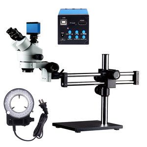
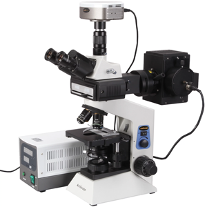
About confocal
Confocal microscopes are advanced optical imaging devices that provide high-resolution, three-dimensional (3D) images of specimens. These microscopes employ a focused beam of light to scan samples, capturing images at different depths and reconstructing a detailed 3D representation. The confocal imaging technique enables researchers to visualize biological samples, semiconductor materials, and other specimens with exceptional clarity and precision. Confocal microscopy finds applications in various fields, including life sciences, material science, and semiconductor research.
How does confocal microscopy work?
In confocal scanning, the microscope uses a focused laser beam to illuminate a single point on the specimen. The emitted light from this point passes through a pinhole aperture that allows only in-focus light to reach the detector. By sequentially scanning multiple points across the specimen, the microscope generates a series of 2D optical sections at different depths. These sections are then compiled to construct a detailed 3D image of the sample. The ability to eliminate out-of-focus light and precisely control the focal plane contributes to the enhanced optical sectioning and depth resolution characteristic of confocal microscopy.
Applications of confocal microscopy
Confocal microscopy is widely used in various scientific disciplines, including cell biology, neuroscience, material science, and microbiology, to visualize and analyze samples at high resolution. In cell biology, researchers can examine the internal structures of cells and tissues with exceptional clarity, enabling detailed studies of cellular processes. Neuroscientists use confocal microscopy to investigate intricate neural networks in the brain and analyze the distribution of specific molecules. The technology is also valuable in material science for imaging semiconductor devices, polymers, and other materials with precision. In microbiology, confocal microscopy aids in studying microbial communities and biofilms.
Confocal microscopy vs conventional microscopy
One of the key differences between confocal and conventional microscopy lies in the imaging technique. Traditional microscopes capture the entire sample thickness at once, resulting in images with less optical clarity, especially in thick specimens. In contrast, confocal laser scanning microscopy employs a focused laser beam and pinhole aperture to eliminate out-of-focus light, enabling the acquisition of optical sections at different depths. This optical sectioning capability reduces background noise and improves the overall image quality in confocal microscopy. Additionally, confocal microscopy allows for the visualization of samples in 3D, providing detailed information about the spatial organization of structures within the specimen. The ability to generate 3D reconstructions is particularly valuable for analyzing complex biological samples and investigating the spatial relationships between different cellular components.