All categories
Featured selections
Trade Assurance
Buyer Central
Help Center
Get the app
Become a supplier
(779 products available)


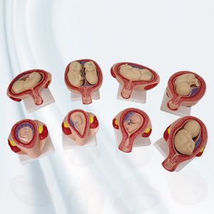


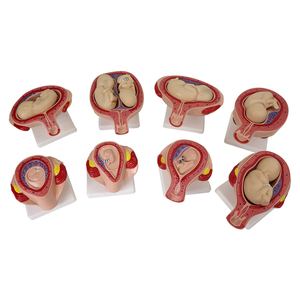


Uterus 3D models are valuable educational tools for medical professionals and students. Thanks to 3D printing and digital modeling, these models provide an intricate and precise view of the uterus and its associated structures like never before. Below are some of the many 3D uterus models available:
These models focus primarily on the anatomical and structural components of the uterus. They depict the muscular walls, endometrial lining, cervix, fallopian tubes, and ovaries. Such detailed representations help in:
Beyond the normal anatomy, some models highlight specific uterine pathologies. Whether it is fibroids, endometriosis, polyps, or uterine septum, these models are essential for educational purposes. These models come in handy during training to:
With advancements in digital technology, interactive 3D models have come into play. The real beauty of these models lies in their:
Due to their versatility, they are widely used in:
These models provide an all-encompassing view of the female reproductive system. It includes not just the uterus but also:
Such comprehensive models assist in:
The design of a 3D model of the uterus is crucial if the model is to serve its intended purpose well and effectively. Various elements make the model, including the software and materials used to make them. Below are the design elements that must be considered when making a 3D uterus model:
The accuracy of a 3D model largely depends on the data used to create it. Medical imaging modalities, including MRI and CT scans, serve as valuable resources for model creators. Only experienced professionals can translate these data forms into accurate models. The accuracy of the model is important because:
Although digital models are gaining popularity, 3D printing has distinct advantages. Among them is the anatomical model for real-life applications, especially for low-tech environments. \These physical models help doctors without much access to hospitals and medical training schools.
However, 3D printed materials vary, and some of the most common include:
Once printed, the models often undergo post-processing. Sanding, painting, and sealing enhance the model’s look and feel.
More importantly, they affect the model’s educational value. For instance, coloring certain areas can help distinguish the endometrium from muscle tissue.
Virtual models are designed using robust software. Common tools include:
These tools give the designer control over the model’s geometry and internal organ characteristics.
3D uterus models serve multiple purposes, each necessitating a different type of model. They go even further and serve a myriad of scenarios. They help in clinical settings to educational systems and beyond. Below are some of the many uses and scenarios of 3D uterus models:
Medical students use models like 3D printed organs to learn vital surgical skills. Hands-on practice on these models boosts their confidence. The confidence boost aids them in performing actual procedures later in their careers.
Furthermore, these models stand out among other training modalities. They determine where key structures such as blood vessels and ligaments are located. This feature helps make the training as close to real-life conditions as possible.
Before going into the operating room, surgeons need to know as much as possible about their patients. This understanding helps prevent mistakes and improve prognosis. That is why they use 3D models to create unique uterine diagrams for each patient.
By examining the model, surgeons can visualize the surgical area. This way, they fine-tune their approaches and lapse any potential complications.
Patients are often confused by medical jargon. This circumstance makes it challenging for them to fully understand their conditions. They also fail to grasp how proposed interventions will help. Doctors use 3D models to bridge this gap.
The physical reproduction helps translate complex ideas into something tangible. It may not always be the case, but using models improves comprehension. It also spares the doctor from re-explaining concepts multiple times.
3D models are also vital in biomedical research. They make it easier for researchers to understand how different conditions affect uterine structure. Researchers also use them to test new treatments, procedures, or medical devices in a controlled setting.
This testing accelerates innovation within healthcare. It is worth noting that customized models also push the quality of research up a notch. They are made to mimic specific pathological conditions or anatomical variations. This feature helps in getting more targeted results and data.
3D models aid medical device developers in testing their inventions. The developers use them to understand how new instruments interact with uterine tissue. This understanding ovulates much-needed safety and efficacy data before commercial launch.
This testing can identify design flaws early, reducing the need for costly modifications later.
3D uterus models can be either digitally stored or physically printed. Regardless of the model type, they require proper care to function optimally. Luckily, spouting and caring for them is pretty easy and straightforward. That said, the following are some of the maintenance and specifications of the 3D uterus models:
3D models come in various formats. They are usually stored in digital healthcare information systems. Such systems include:
Databases store digital models safely, securely, and accessibly. Users can access them right from their workstations. However, they must have the correct access permissions to access them.
On the other hand, physical models require more tangible storage solutions. Usually, they should be kept in protective cases to avoid damage. The cases further ensure the models remain clean and ready for usage.
Digital models require little maintenance. Users need to back them up to avoid loss. The models should also be updated occasionally. This update ensures the model incorporates the latest anatomical data or technological advancements.
Hospitals and training institutions also use scanners and printers to create digital copies of physical 3D models. They then archive the models on a computer and in the cloud, just like digital models, to avoid losing them.
Fortunately, maintaining physical models is not that hard either. Simply cleaning them regularly keeps them in top condition. Models made from plastics, metals, or resins are generally easy to wipe down.
Meanwhile, models made from more delicate materials like 3D printed plastic require gentler cleaning methods. Gentle practices ensure they do not lose detail and remain hygienic for clinical use.
Digital models enjoy excellent longevity. As long as they are backed up, they can be accessed indefinitely. In fact, they can be modified or updated as medical technology progresses. This progress ensures that the model remains relevant for years to come.
Conversely, physical models have a shorter lifespan. Factors such as wear, tear, and environmental conditions degrade them. Nevertheless, with proper care, they can still offer several years of reliable service.
Version control is vital in maintaining digital 3D models. As medical knowledge evolves, models may need updates. Tools like GitHub for 3D files provide version control.
The tools track changes, making it easy to revert to earlier versions if necessary. Conversely, physical models do not have version control. They can be modified only through physical means and methods.
A1. Plastic filaments like PLA and ABS are the most commonly used materials. Hospitals also use resin for more detailed models and durable metals like stainless steel.
A2. One of the joys of digital 3D models is their reusability. As long as the digital files are securely stored, they can be reproduced, modified, and shared as often as needed.
A3. Doctors use 3D models to demonstrate complex medical concepts. Doing this enables them to bridge the gap between layman terms and medical jargon. This bridge enhances patient understanding, making patients feel more involved in their treatment journey.
A4. Yes, 3D printed human organs that will be placed inside patients’ bodies have to be sterilized. Commonly used sterilization methods include:
A5. Medical imaging software like OsiriX is popular for creating 3D models for printing. Other common tools include Blender, Fusion 360, and ZBrush. Hospitals and medical institutions prefer Fusion 360, as it is easy to use.