All categories
Featured selections
Trade Assurance
Buyer Central
Help Center
Get the app
Become a supplier
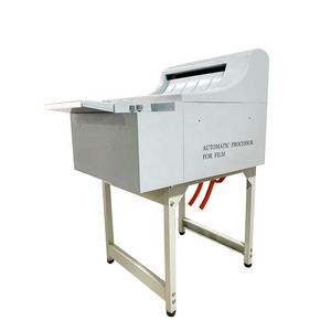
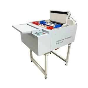
(858 products available)


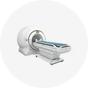
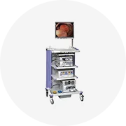
Digital image processing in medical devices and supplies is used to enhance x-rays and, therefore, the images doctors use to diagnose their patients. Several types of technology can process these images, and each serves a different purpose depending on the patient's medical need.
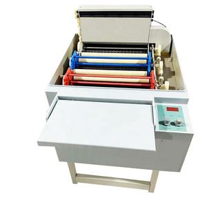
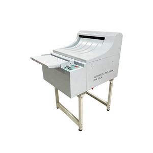
Early systems used merely analogue technology to process x-ray images. However, these early systems employed hardware to aid in processing; for instance, the edge-enhancement filter was a rudimentary analogue filter mounted on an x-ray tube envelope, designed to improve the resolution of the x-ray images by heightening some specific details and contours.
Circuit boards with custom filters for contrast augmentation of frames and other processing were widely used in older systems. They were retrofitted to conventional x-ray equipment in two ways: directly between the generator and the x-ray tube or remotely connected to the detector and x-ray source.
As x-ray systems moved from film to digital imaging, most of the image processing systems got migrated to workstations for clinical use. These workstations are utilised to pre-process images prior to any diagnostic manipulation.
Medical x-ray equipment manufacturers usually build these workstations. In radiology, image processing is often provided via an optional mount for the workstation, such as filtering, transformation, contrast, and threshold. Filters used in image processing encompass spatial filters to rectify noise, define edges, and improve the image's overall appearance.
Post-processing is considered an essential processing step, typically after the x-ray examination or before the x-ray image becomes integrated into the patient's medical record and viewed by the physician. A general workstation that solely focuses on image processing is often used to conduct post-processing.
This processing enhances the image for clarification so that the doctor can see an improved version when viewing the picture. Post-processing involves operations like contrast stretching to use the entire intensity range, edge enhancement to use structural details, and area of interest to eliminate shadows and brightness to improve the image quality further.
Post-processing is often integrated into PACS (Picture Archiving and Communication System) software. Still, it can also be performed independently using third-party software, which may not be as effective as integrated processing but can be cost-effective. Manufacturers and imaging centers frequently create customized filters and enhancement algorithms for particular modalities and imaging circumstances.
X-rays are essential tools that offer a view of the internal structures of a living body in modern medicine, and image processing plays a vital role in enhancing the clarity and utility of these images.
This is majorly concerned with improving the features of an x-ray image, such as the resolution and visibility of the underlying details. After making an x-ray, several techniques are employed to improve the image, such as rectifying noise, improving the contrast, and clarifying the minute details captured in the picture. For example, spatial filtering acts as a noise reduction tool to remove the noise present in the image, while histogram equalization is a way to improve contrast through equalisation of the histogram and optimisation of the image's brightness levels.
Commonly known as: It refers to the usage of available image data to create or reconstruct a picture that will affect specific internal body parts. Advanced machine learning techniques, such as deep learning, generate new images from the existing volumetric dataset. They retrieve or create internal views of organs and tissues in three-dimensional. These images can also be used for surgical planning by providing accurate-size images of internal organs or parts of the body.
Medical x-ray image processing uses digital techniques to improve image formation and quality. Apart from storage and transmission of the images, digital systems have various advantages over earlier systems, such as faster image retrieval and greater accessibility for healthcare professionals. Furthermore, a feature of digital x-ray systems is the storage and transmission of x-ray images. Using compressed image formats aids users in storing their images without using much space and transmitting them quickly through networks. X-rays can be digitised in PACS for efficient transfer over the internet to facilitate remote consultations by keeping in mind the confidentiality and security of personal health data through encryption.
Image analysis is concerned with performing automatic image analysis, such as identifying or segmenting certain features in the x-ray, such as lesions, tumours or infections. Various advanced AI systems have been developed over the years and are currently being used in radiology to help doctors quickly and accurately analyse their x-ray pictures. The radiologists are assisted through automated image analysis in detecting abnormalities, thus resulting in improved diagnostic accuracy and consistency of the diagnosis across different patients.
The commercial value of digital X-ray image processing is in its contribution to improved medical diagnostics, and the use is primarily in the healthcare sector, especially radiology and orthopaedics, for better disease detection. It helps reduce unnecessary surgical interventions benefiting both healthcare providers and patients, so many healthcare centres invest in these systems.
Cost and pricing considerations
Pricing for x-ray image processing systems is dependent on their features, processing power, and the software's complexity algorithms. Advanced systems endorsed with artificial intelligence offer superior diagnostic services and may attract higher investment costs. The budget and the volume of patients handled daily determine the healthcare centre or imaging center's pricing system. PACS is implemented by large hospitals and radiology practices and is a well-featured and expensive system, while standalone systems for processing images can be used by smaller clinics and imaging centers, which are relatively cheap.
Return on investment (ROI)
The ROI of digital x-ray image processing systems is observed through better operational effectiveness, improved patient outcomes, and increased revenues resulting from handling more diagnostic services. High-resolution imaging coupled with advanced image processing leads to improved accuracy in disease diagnosis. Thus, this will cause the decrease in treatment costs due to fewer repeat tests or incorrect diagnoses. These systems also increase revenue for healthcare facilities by increasing patient volumes and providing more services required for effective diagnosis and treatment.
Market trends
Various trends keep on happening in the x-ray image processing market, such as artificial intelligence and telemedicine. Image processing with AI, deep learning inclusive, is revolutionising x-ray interpretation, leading to faster and more accurate diagnosis. Another compelling aspect is the need for cloud-based x-ray imaging solutions due to the increasing workload and need for outside consultation in healthcare. They facilitate efficient collaboration by enabling remote access to images and reports. Mobile applications for x-ray interpretation and tele-radiology are gaining importance and positively impacting radiology practice.
Regulatory considerations
The FDA regulations must be adhered to while considering the advantages of digital x-ray image processing. Digital x-ray systems and image processing software must go through regulatory approval to satisfy standards of safety and efficacy.
Several factors are taken into consideration when choosing a system for processing x-ray images digitally.
Technical specifications
Technical specifications also affect the choice of a digital x-ray system, one of which is resolution, as higher resolution translates into clearer images; thus, the detail will be more visible. The x-ray machine should cover the modality in the imaging centre, such as radiography or fluoroscopy. Processing time is also vital, as shorter times will enhance the system's efficiency. Special attention should be paid to the software features involved, such as filtering, contrast adjustment, and enhancement.
Budget
Financial plays a vital role in deciding which system to choose. Standalone systems offer digital processing at a relatively low price and can be easily afforded by small clinics and imaging centres. More advanced systems like PACS can be relevantly expensive, but they facilitate considerable efficiency and collaboration, ROI, and operational effectiveness.
Integration and compatibility
The chosen x-ray processing system must be compatible with other equipment used in imaging centres or hospitals, such as his RIS (Radiology Information System) and PACS. The selected system should also be integrated into existing workflows. Tele-radiology solutions rely heavily on this integration since they are designed to facilitate remote consultations.
Scalability
The imaging facilities should select an x-ray processing solution that will be able to accommodate future growth, especially when there is an increase in the volume of patients or when new modalities are needed. Systems with flexible architecture allow for enhancement or adjustment in the business as it expands or changes. Cloud-based solutions in imaging centres are a good example of scalable options, as they provide the necessary resources to handle increased workloads and facilitate a decent degree of accessibility.
Maintenance and support
Adequate technical support and low-maintenance requirements go a long way in ensuring that x-ray processing systems have maximum uptime. It would be a good idea to research the level of assistance provided by the vendors and the ease with which potential problems can be addressed. The total cost of ownership, which includes maintenance and support over time, should be factored in when developing a budget for the project.
Here are frequently asked questions about digital x-ray image processing.
Q1: What is digital x-ray image processing?
A1: Digital x-ray image processing refers to improving x-ray images through advanced techniques like filtering, enhancing, and analysing the pictures to make them clearer and more useful for making medical diagnoses.
Q2. What are the benefits of digital x-ray image processing?
A2. X-ray images can be stored, shared, and retrieved more easily. They also enable improvements, thus providing better icons of that image that may lead to better diagnostic accuracy and detail recognition.
Q3. How does image processing enhance the quality of x-ray images?
A3. Methods in post-processing like noise removal, contrast improvement, and edge enhancement work together to improve the quality of the picture and the clarity of internal structures in patients.
Q4. What tools or software are commonly used for x-ray image processing?
A4. Filters, software, and workstations are used to pre-process x-ray images, including third-party software and custom filters integrated into the PACS and algorithms from various manufacturers.
Q5. Is digital x-ray image processing expensive?
A5. The expense will depend on several factors, such as the system's features, the hospital's or imaging centre's budget, and the volume of patients handled daily. Standalone systems are affordable for smaller facilities, while larger ones, like PACS, may be a significant investment.