All categories
Featured selections
Trade Assurance
Buyer Central
Help Center
Get the app
Become a supplier
(312 products available)


 Ready to Ship


 Ready to Ship


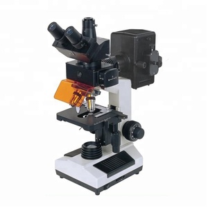
There are several types of fluorescence microscopes with cameras, each suited for specific applications and requirements. These microscopes combine the structural details of traditional light microscopy with the functional and molecular insights provided by fluorescent microscopy. Here's a breakdown of the main types:
Widefield Fluorescence Microscope
The structure of a widefield fluorescence microscope is simple, and it is often used to observe fluorescently tagged samples. This microscope captures the entire field of view, making it suitable for general fluorescence microscopy applications. However, due to the large amount of out-of-focus light, its resolution is limited.
Confocal Laser Scanning Fluorescence Microscope
This fluorescent microscope is equipped with a laser scanning system that allows users to obtain sharp fluorescence images of samples. Using the confocal technique, this microscope eliminates most of the out-of-focus light to capture clearer images. It captures a fluorescent micrograph of a processed sample while scanning it in thin optical sections.
Super-resolution Fluorescence Microscope
Super-resolution fluorescence microscopes can resolve structures at a nanometric scale, which far exceeds the diffraction limit of conventional fluorescence microscopes. In this type of microscope, techniques like STED, SMLM, and PALM are used to improve spatial resolution and facilitate imaging at a much finer level.
Live-cell Fluorescence Microscope
A live-cell fluorescence microscope is designed for imaging living cells over time. This model of fluorescence microscope uses advanced imaging techniques to minimize phototoxicity and photobleaching, allowing users to monitor dynamic processes in living biological samples.
Multiphoton Fluorescence Microscope
Using a multiphoton approach, fluorescent microscopes enable deep imaging within thick biological tissues. It uses infrared lasers to excite fluorophores through nonlinear two or three-photon absorption. This method reduces scattering and increases tissue penetration, making it ideal for in vivo imaging of live animals or organomes.
Fluorescence Lifetime Imaging Microscope
A fluorescence microscope camera analysis is based on fluorescence lifetime rather than emission intensity. FLIM distinguishes between different fluorophores by measuring their lifetimes, providing additional contrast and functional information. What this device does is add functional contrast to the images by measuring the lifetime of fluorescence.
The applications of a fluorescence microscope with a camera are wide-ranging across various fields. These applications leverage the capability to visualize fluorescently labeled samples with high spatial and temporal resolution. Below are the categories in which this device applies:
Cell Biology
A fluorescence microscope with a camera is mostly used to study cellular organelles, proteins, and molecular interactions. Cell structures are visualized by tagging specific proteins with fluorescent dyes or antibodies. This enables functional studies of components within live or fixed cells to understand cellular mechanisms and signaling pathways.
Tissue Imaging
In histology and pathology, it is used to identify specific cell types, tissues, or disease markers in thin tissue sections. Fluorescent antibodies or probes that bind to specific antigens enable researchers to visualize cellular structures or pathological changes that can help to understand diseases at a molecular level.
Molecular and Protein Dynamics
In this imaging technique, techniques like FRET (Fluorescence Resonance Energy Transfer) are used to study protein-protein interactions, conformational changes, and molecular dynamics in biological systems. This is mainly useful for understanding complex biochemical processes, signaling pathways, and molecular mechanisms in various organisms.
Immunofluorescence
Immunofluorescence microscopy identifies and localizes proteins or antigens within cells or tissues. It uses antibodies conjugated to fluorescent dyes that bind specific targets in biological samples. This method provides spatial information about protein distribution and helps in distinguishing between different cell types or tissue structures.
Microbial Imaging
In microbiology, fluorescent probes and dyes are used to label and visualize bacteria, fungi, or other microorganisms in environmental or clinical samples. This aids in identifying microbial populations, studying their morphology, and observing dynamic processes like cell division or metabolic activity in real time.
Live-cell Imaging
Fluorescence microscopes are especially prepared to minimize phototoxicity, thus allowing the observation of live organisms. It monitors dynamic biological processes such as cellularmovement, division, or differentiation in real time. This technique is especially important for studying complexations like stem cell or developmental biology.
In-Vivo Imaging
External fluorescent probes permit non-invasive imaging of biological processes in living organisms. This is extremely important for tracking cellular therapies, visualizing tumor growth, or understanding disease mechanisms. It provides insights into the spatiotemporal dynamics of molecular interactions within intact biological systems.
Quality Control and Materials Science
A fluorescence microscope with a camera can also be useful for visualizing defects, chemical distributions, or structural properties in materials science. In this case, it identifies fluorescent markers or tags within composite materials or structured polymers. This technique enhances quality control processes by detecting contaminants, irregularities, or defects in industrial materials.
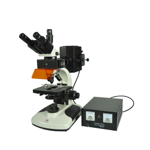
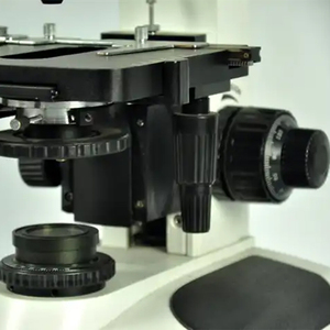
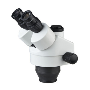
Optical System
The optical system of this type of microscope consists of an objective lens, condenser, and eyepiece. These components work together to capture the fluorescent light emitted by the sample. Widefield fluorescence microscopes use standard optics, while advanced techniques like confocal require lasers and spatial filters to enhance resolution.
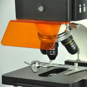
Fluorescence Filters
Fluorescence filters are crucial components of a fluorescence microscope that separates the emitted fluorescent light from the excitation light. These filters, including bandpass and dichroic filters, enable the detection of specific fluorescent signals corresponding to different probes or dyes used in the imaging.
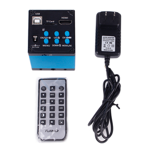
Camera Specifications
A fluorescence microscope camera is used to capture high-resolution fluorescent images. Important camera specifications include resolution, sensitivity, and frame rate. Fluorescent Live Cell Imaging usually requires a high-resolution camera to capture detailed images, while quick cellular dynamics observation favors a high frame rate for capturing rapid processes.
Imaging Software
This technique usually comes with advanced imaging software for photo capturing, photo processing, and analysis. This software facilitates camera operation, fluorescence intensity quantification, and image enhancement. Some of them may also offer features for three-dimensional or time-lapse imaging tailored for specific research needs.
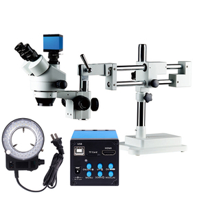
The fluorescence microscope camera attaches to a fluorescence microscope to capture and record high-quality images and videos of fluorescently labeled samples. The microscope is first prepared in the usual way by placing the fluorescent probe on the stage and focusing using the appropriate objective lens. The camera is then connected to the microscope, either via USB or a C-mount, depending on the type of microscope and camera. The camera settings, including exposure time, gain, and resolution, are then adjusted to obtain sharp and well-exposed images. The fluorescence filters should also be selected to match the probe or dye, and the software on the computer connected to the camera should be activated to view and capture live images from the fluorescent microscope with camera.
Good maintenance practices ensure the long-term viability and optimum workings of a fluorescence microscope camera. Here are some of the kinds of maintenance and repair that can be done:
Maintenance
Regular cleaning of both the camera and the fluorescence microscope is important in maintenance. For the camera, the lens should be cleaned using proper lens tissue and solutions to avoid image contamination. Also, the sensor should be checked regularly and cleaned off dust with a blower. For the fluorescence microscope, the regular cleaning of objectives and stages will prevent contamination and allow smooth functioning. Also, regular checking of the filter sets, wavelength check, and bulbing of lasers will ensure proper imaging. Proper storage of both the camera and fluorescence microscope when not in use will protect them from dust and accidents.
Repairs
If the fluorescence microscope camera begins to show problems, troubleshooting procedures can be employed before considering repairs. For the camera, basic troubleshooting includes checking settings or connections. For the fluorescence microscope, basic microscopy troubleshooting includes checking the bulb and adjustment of the focus. However, complex problems, such as internal damage, calibration issues, or malfunctioning parts, usually require expertise for repair. The repair may include the replacement of internal parts of the camera, such as the sensor or circuit board. The repair of the fluorescence microscope may entail replacing worn objective lenses or repairing the laser system.
For effective performance, several quality and safety factors need to be considered when selecting fluorescent microscopes with camera systems. These factors ensure reliable imaging, optimal sample protection, and user safety.
Fluorescence Microscope Camera Quality
Fluorescence microscope camera quality is one of the most important factors to consider since poor quality will lead to poor results. A high-resolution camera, such as a digital EM-CCD or sCMOS camera, will ensure great-resolution images needed for fine morphological studies. Fluorescence sensitivity and low-noise are also important factors for capturing dim signals without loss of quality.
Microscope Quality
The fluorescent microscope itself should have well-corrected objectives and a wide-field aperture to capture maximum fluorescence. Using quality filters and dichroic mirrors enables better separation of excitation and emission wavelengths. Also, relying on manufacturers with a reputation for confocal and widefield fluorescence microscope technology is mandatory for quality checks and after-sales services.
Sample Preservation
To facilitate quality imaging, special attention should be paid to minimizing phototoxicity and photobleaching, especially with biological samples. Using immersion oil reduces phototoxicity of living cells, and low-power lasers minimize photobleaching in vulnerable samples. Rapid imaging and exposure times are employed to reduce light intensity on sensitive samples to avoid damage during immediate imaging.
Laser Safety
Working with a laser confocal and other advanced fluorescence microscopes incurs caution and proper handling. These lasers must be handled with care to prevent damage to the eyes, so microscopes come with proper protective covers and ports, and these should be capped and guarded properly. Working personnel must also be trained on proper handling and exposure limitation before operating laser devices.
Chemical Safety
Fluorescent probes, dyes, and antibodies may contain hazardous chemicals. Adequate personal protection equipment (PPE), such as gloves and safety glasses, are a must when handling these reagents. Proper storage and disposal procedures must also be ensured to prevent accidents and environmental contamination.
Equipment Safety
To ensure equipment safety, appropriate handling practices for both the fluorescent microscope and the camera must be ensured. This includes regular checking and maintenance of the system to prevent malfunction, which may cause accidents. Moreover, all equipment must be properly grounded and shielded to avoid electrical accidents, especially when operating computer interfaces with cameras attached to microscopes.
A1. Fluorescence microscopes with cameras are mainly used to study biological samples, tissues, and cells tagged with fluorophores. They allow visualization and imaging of the fluorescent signals emitted by these samples, providing detailed information about structures and dynamics at the microscopic level.
A2. A fluorescence microscope combines conventional optical microscopy with fluorescent imaging. This allows the user to visualize and capture fluorescently labeled specimens. Using specific wavelengths of light, the microscope excites fluorophores within the samples to emit light, which the camera captures to create images showing the distribution and interaction of Target molecules.
A3. Fluorescence microscopes are particularly useful for visualizing specific proteins, cells, or structures within a complex biological context. It provides the ability to examine dynamic processes in living cells and tissues, offering insights into cellular mechanisms, disease progression, and drug effects at an unparalleled level of detail.
A4. A fluorescence microscope typically consists of an optical system, fluorescence filters, and a camera. The optical system includes an objective lens and condenser that capture the fluorescent light emitted by the sample, while the filters separate the excitation and emission wavelengths, and the camera would capture and analyze the images.
A5. To maintain a fluorescence microscope with a camera, users should clean the lenses and filters regularly, avoid harsh chemicals, and use proper lens tissue to prevent damage. Also, the camera sensor should be checked and dusted using a blower, and both the camera and microscope should be stored in a protective environment when not in use.